

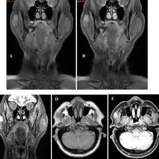

Neuroradiology uses specialized imaging techniques to evaluate conditions affecting the brain, spine, head, neck, and nervous system. Radiologists use technologies such as MRI and CT imaging to produce detailed views of neurological structures and abnormalities. These images provide physicians with information that supports diagnosis, treatment planning, and ongoing evaluation of neurological conditions. Here is more information about the advances in neuroradiology research and clinical applications:
Using Advanced Diagnostic Imaging
Neuroradiology uses advanced imaging technology to evaluate abnormalities affecting the brain, spine, head, neck, and nervous system. Specialized MRI and CT techniques provide detailed images that help radiologists identify structural changes and other neurological findings. These images provide information that physicians use when evaluating symptoms and developing treatment plans.
Radiologists interpret imaging studies alongside medical history, symptoms, and other clinical information. Advanced imaging methods provide additional detail when evaluating complex neurological conditions or changes requiring further examination. Physicians use these findings to guide additional testing, treatment decisions, and ongoing neurological care.
Using Advanced MRI Techniques
Advanced MRI techniques provide detailed information about brain structure, function, and neurological activity. Diffusion tensor imaging maps white matter pathways within the brain and provides information about nerve fiber organization. Physicians may use these images when evaluating neurological conditions or planning certain procedures.
Functional MRI measures changes in blood flow associated with activity in different areas of the brain. This imaging technique helps identify regions involved in functions such as movement, speech, and cognition. Neurosurgical teams may use this information when planning procedures near areas responsible for these functions.
MR spectroscopy provides information about chemical activity within tissues rather than displaying structural anatomy alone. These findings can supplement conventional MRI when physicians evaluate certain brain abnormalities and neurological conditions. Neuroradiologists select imaging techniques according to the condition, clinical question, and information needed for further evaluation.
Performing Minimally Invasive Neurointerventions
Interventional neuroradiology uses imaging guidance to diagnose and treat certain conditions affecting blood vessels within the brain. Cerebral angiography provides detailed images of blood vessels when evaluating aneurysms, vascular malformations, and other neurological conditions. Specialists also use fluoroscopic imaging during procedures such as mechanical thrombectomy to locate and remove certain blood clots. Imaging guidance also supports treatments involving embolization and flow-diverting devices for selected cerebral aneurysms.
Advancing Spinal Imaging
Spinal neuroradiology uses specialized imaging techniques to evaluate the spinal cord, nerve roots, discs, and surrounding structures. Different techniques provide information based on symptoms, previous procedures, and individual imaging needs. Available spinal imaging approaches include:
- Dynamic MRI for evaluating spinal structures and changes during movement
- Myelography for examining the spinal cord, nerve roots, and surrounding spaces
- CT myelography for providing detailed images when standard MRI presents limitations
- Quantitative MRI for evaluating tissue characteristics within spinal discs and the spinal cord
Neuroradiologists select imaging methods according to the suspected condition, clinical findings, and information needed for further evaluation.
Advancing Stroke and Vascular Assessment
Neuroradiology uses specialized imaging techniques to evaluate blood flow and vascular changes within the brain. CT and MR perfusion imaging provide information about brain tissue affected by reduced blood flow during certain strokes. Vessel wall MRI provides detailed images of blood vessel walls and abnormalities that standard angiography may not fully characterize. Additional techniques, including advanced flow imaging, provide information about blood movement within cerebral vessels during neurovascular evaluation.
Schedule Your Neuroradiology Appointment
Neuroradiology uses advanced imaging to evaluate conditions affecting the brain, spine, nervous system, and surrounding blood vessels. Available examinations include brain and spine MRI, head CT, cerebral angiography, and myelography based on individual diagnostic needs. These imaging studies provide detailed information that physicians use when evaluating neurological symptoms and planning further care. Schedule a neuroradiology appointment today to discuss your imaging needs and appropriate diagnostic options.
- The Benefits of Seeing a Back Doctor for Sports Injuries
- How Skin Removal Surgery Helps Enhance Weight Loss
- How Industrial Teams Can Prevent Costly Equipment Downtime
- A Complete Guide To Filing A Medical Malpractice Claim In Georgia
- Find an Inclusive Therapist in Chicago, Illinois: Culturally Responsive Care With Ida Lillie Psychotherapy